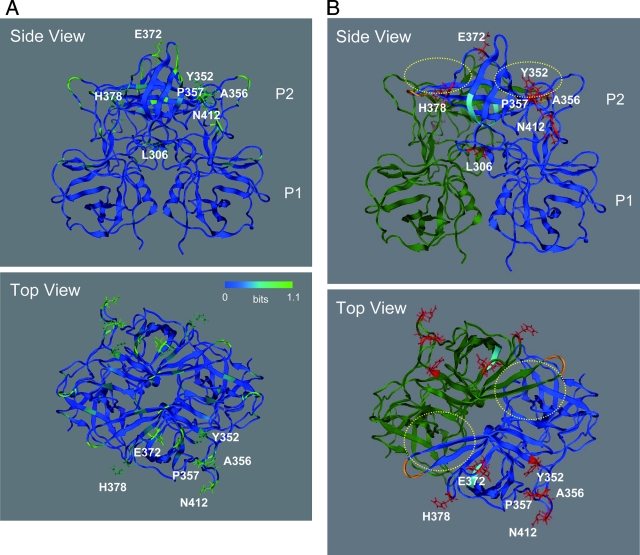
FIG. 6.
Structural model of the VP1 P domain dimer of the NoV GII/4 2006b strain. The model was constructed by homology modeling using the X-ray crystal structure of the P domain dimer of the 1995-1996 epidemic GII/4 strain (4). (A) Shannon entropy scores expressed on the P domain model. (B) Side and top views of the P domain model. Reported functional sites for virus entry into the cells are highlighted. Yellow dot circles, the fucose ring binding sites formed by P-domain dimer (4); cyan chain, an RGD motif (48) on the β2 sheet of the P domain; orange chain indicates an additional RGD-like motif, KGD (46), on the tip of the β4-β5 loop of the P domain. Red sticks indicate side chains of amino acids unique to the 2006b strains.

