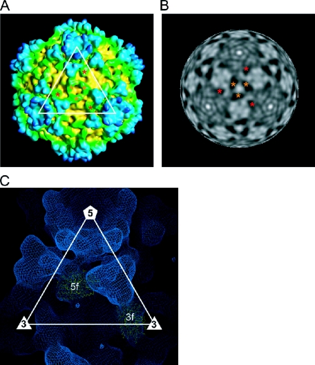
FIG. 4.
Clamp proteins. (A and B) Surface representation (A) and radially cued density at a radius of 293 Å (B), viewed along a threefold axis. Two icosahedrally independent 3f and 5f clamp proteins are marked with orange and red asterisks, respectively. (C) The atomic structures of the clamp σ2 protein of Orthoreovirus were fitted into the cryo-EM map of RRSV.

