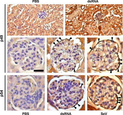
FIG. 5.
Induction of p49 and p54 in the glomeruli of murine kidneys. At 8 h after i.v. injection of dsRNA, SeV, or PBS, mice were sacrificed, kidneys were collected, fixed, and processed for immunohistochemistry to detect p49 (upper and middle panel) and p54 (lower panel). White arrowheads indicate mesangial cells, black arrowheads indicate podocytes in the blood-filtrating glomeruli, located in the renal cortex. Magnifications are ×200 (upper panel) and ×400 (middle and lower panels). Scale bar, 25 μm.

