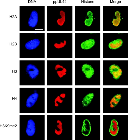
FIG. 2.
Subnuclear distribution of core histones in relation to late viral replication compartments. MRC-5 cells were infected with CMV for 48 h, fixed with paraformaldehyde/methanol, and stained with a mouse monoclonal antibody specifically detecting the CMV ppUL44 DNA polymerase accessory protein and rabbit polyclonal antibodies directed against the C-terminal histone fold domain of H2A, H2B, H3, or H4. A rabbit antiserum specific for histone H3 dimethylated at lysine 9 (H3K9me2) was used as a negative control. Samples were subsequently stained with DAPI, a mouse-specific Alexa Fluor 594, and a rabbit-specific Alexa Fluor 488 conjugate. Representative nuclei showing DAPI, ppUL44, and the respective histone staining are shown. Additionally, merge images of ppUL44 and histone signals are presented. Scale bar, 10 μm.

