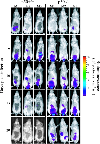
FIG. 2.
Mice lacking the p50 subunit of NF-κB show prolonged carriage of C. rodentium in the abdominal region. In vivo colonization and clearance dynamics were monitored by BLI in p50+/+ control mice (left) and p50−/− mice (right) during C. rodentium infection. The images were acquired using an IVIS50 system and are displayed as pseudocolor images of peak bioluminescence, with variations in color representing the light intensity at a given location. Red represents the most intense light emission, while blue corresponds to the weakest signal. The color bar indicates relative signal intensity. The mice were imaged at various time points p.i., with an integration time of 1 min. Three representative p50+/+ control animals and three representative p50−/− animals are shown.

