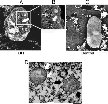
FIG. 2.
LKT on mitochondria isolated from BL-3 cells detected by scanning electron microscopy and transmission electron microscopy. Mitochondria isolated from BL-3 cells were incubated with 0.5 U LKT (A and B) or RPMI medium (control) (C), stained with anti-LKT colloidal gold, and visualized by scanning electron microscopy. The micrographs show areas of LKT binding (clusters of gold beads in panels A and B) and sloughing of the MOM, leaving punched-out areas (arrows in panels A and B). (D) Mitochondrion with a disrupted outer membrane and cristae (black arrows) and normal mitochondrion with an intact double membrane (white arrow). Bars = 200 nm.

