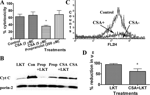
FIG. 4.
CSA protects BL-3 cells against LKT-mediated cytotoxicity, mitochondrial membrane damage, and collapse of ψm. (A) BL-3 cells (106 cells) were preincubated with CSA (2 or 5 μM) for 30 min before incubation with LKT (0.5 U) for 60 min. Cytotoxicity was measured using the Cell Titre 96 AQ one-assay system. (B) Mitochondria were immunopurified from untreated BL-3 cells, preincubated with CSA (5 μM) or with propranolol (Prop) (200 μM), and then incubated with LKT (0.2 U) for 30 min at 37°C. Immunoblot analysis of the resulting mitochondrial lysates demonstrated that CSA, but not propranolol, protected against LKT-mediated mitochondrial cytochrome c (Cyt c) release. Con, control. (C and D) BL-3 cells were preincubated with CSA or medium (control) and then incubated with LKT (0.5 U) for 1 h. Cells were then incubated with JC-1, a ψm-sensitive dye, at 37°C for 15 min and washed three times in PBS. Aggregated intramitochondrial JC-1 is red, and the monomeric cytoplasmic form is green. BL-3 cells were then analyzed by flow cytometry in the FL2 channel (absorption and emission maxima at 580 and 595 nm, respectively). The results show that CSA protected against collapse of ψm. The bars in panel D indicate the means of three separate experiments, and the error bars indicate the standard errors of the means. *, P < 0.05.

