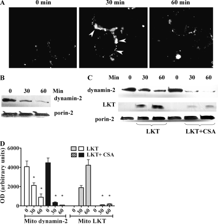
FIG. 5.
Movement of dynamin-2 to the cell periphery in LKT-treated BL-3 cells. (A) BL-3 cells (106 cells) were incubated with LKT (0.5 U) for 30 or 60 min and then fixed, permeabilized, stained for cytoplasmic dynamin-2 (arrows), and visualized by confocal microscopy. (B) BL-3 cells were incubated with CSA (5 μM for 30 or 60 min at 37°C). Mitochondria were isolated and lysed, and the lysates were immunoblotted for dynamin-2 and porin-2. (C) BL-3 cells preincubated with CSA were treated with LKT for 30 or 60 min. Their mitochondria were then isolated and lysed, and the lysates were immunoblotted for LKT, dynamin-2, and porin-2. (D) Mitochondrial (Mito) dynamin-2 and LKT levels in immunoblots as determined by densitometry. The bars indicate the means of three separate experiments, and the error bars indicate the standard errors of the means (*, P < 0.05). OD, optical density.

