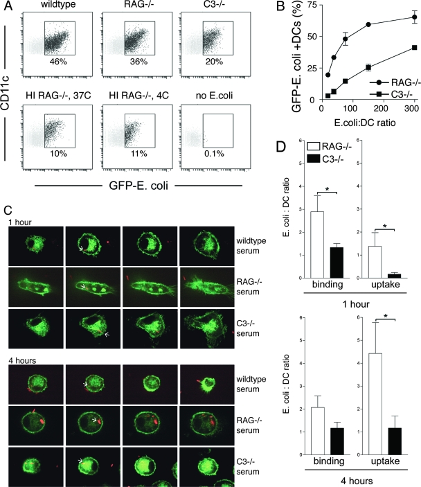
FIG. 1.
C3-derived opsonins and Ig facilitate phagocytosis by mouse DCs. (A) Fresh sera from wild-type, RAG1−/−, and C3−/− mice, as well as heat-inactivated (HI) serum from RAG1−/− mice, were used to opsonize GFP-expressing E. coli. Wild-type serum contains complement and Ig; RAG1−/− serum is complement sufficient but Ig deficient; C3−/− serum lacks complement C3 but is sufficient in Ig; heat-inactivated RAG1−/− serum is deficient in both complement and Ig. GFP fluorescence was measured in DCs after 60 min of internalization using an MOI of 50:1. (B) GFP-expressing E. coli bacteria were opsonized using fresh sera from RAG1−/− and C3−/− mice, and titers were determined on DCs in twofold dilutions (MOIs of 300:1, 150:1, 75:1, 38:1, and 19:1). RAG−/− serum promoted superior E. coli phagocytosis at all MOIs. Data are representative of more than five independent experiments. (C) Opsonized DsRed-E. coli bacteria were added to DCs derived from class II-EGFP mice and analyzed for deposition into class II-EGFP-positive endosomal compartments, using class II-EGFP as a marker for late endosomes/lysosomes (7). Four consecutive Z-stack sections are shown through the center portion of DCs downward (farthest-right images show dendrites attached to the coverslip). White arrows point toward endosome-localized DsRed-E. coli. (D) Thirty fields were analyzed by Z-stack analysis, each visualizing several DCs (×100 magnification; at 1 and 4 hours of DC culture in the presence of DsRed-E. coli), and E. coli binding and uptake by DCs were enumerated. *, statistically significant at P < 0.05.

