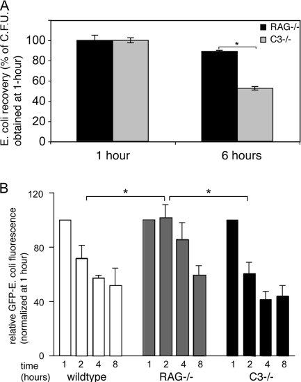
FIG. 2.
Ig facilitate phagosomal degradation of E. coli antigen. (A) Opsonized E. coli bacteria were added to DCs for 1 or 6 hours, after which gentamicin was added to kill remaining extracellular E. coli bacteria. DCs were resuspended and plated in serial dilutions and in triplicate onto LB-agar plates. CFU from endocytosed E. coli were expressed as a fraction of the number recovered after 1 hour of incubation. (B) Live opsonized GFP-E. coli bacteria were added to DCs for 1, 2, 4, and 8 hours, after which DC uptake and processing were stopped and GFP fluorescence in DCs was determined by flow cytometry by counterstaining for the DC marker CD11c. GFP fluorescence at 2, 4, and 8 hours was plotted as a percentage of GFP fluorescence determined at 1 hour of culture using wild-type, RAG−/−, and C3−/− serum opsonization. *, statistically significant at P < 0.05.

