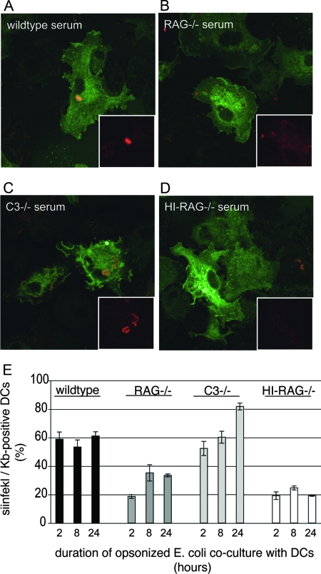
FIG. 4.
Formation of peptide/class I MHC complexes through cross-presentation requires Ig. (A to D) OVA-E. coli bacteria were opsonized with serum from wild-type (A), RAG1−/− (B), or C3−/− (C) mice or with RAG1−/− serum depleted of complement by heat inactivation (HI) (D). Opsonized nonfluorescent E. coli bacteria were then added to MHC class II-EGFP-expressing DCs, and at 2 hours, DCs were fixed and stained for SIINFEKL/Kb complexes using 25D1.16 antibody. Only wild-type and C3−/− serum-opsonized E. coli bacteria were processed sufficiently at this time to detect intracellular SIINFEKL/Kb staining. (E) Surface display of SIINFEKL/Kb complexes was analyzed by flow cytometry after coculture of OVA-E. coli with DCs for 2, 8, and 24 h. E. coli bacteria were opsonized as described for panel A and added to DCs. Opsonization using wild-type and C3−/− sera yields surface display of SIINFEKL/Kb complexes in greater than 50% of DCs starting at 2 hours postuptake. These data show that while RAG1−/− serum promotes phagocytosis, opsonization using RAG1−/− serum does not yield significant SIINFEKL/Kb presentation (20% of DCs express SIINFEKL/Kb complexes at 2 hours postuptake, which is similar to what is found using heat-inactivated RAG−/− serum).

