
FIG. 2.
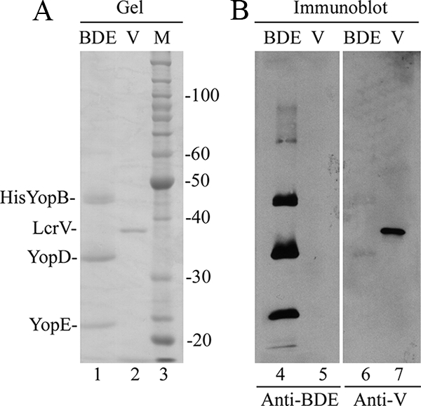
SDS-PAGE and immunoblot analyses of BDE and LcrV. (A) Samples of purified BDE (lane 1) and His-LcrV (V; lane 2) were resolved by SDS-PAGE and visualized in the gel by staining them with GelCode Blue. The positions of His-YopB, LcrV, YopD, and YopE are shown on the left. A molecular weight (MW) standard (M; lane 3) and corresponding MWs are shown on the right. (B) Proteins in gels that duplicate those shown in panel A were transferred to membranes. The membranes were processed by immunoblotting to detect antigens recognized by IgG antibodies in sera from mice immunized with BDE (anti-BDE; lanes 4 and 5) or LcrV (anti-V; lanes 6 and 7).