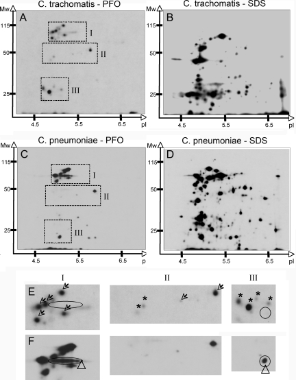
FIG. 4.
Numerous Chlamydia proteins localize within the eukaryotic cell cytosol. Autoradiogram of the soluble fraction obtained from C. trachomatis-infected HeLa cells (A, B, and E) or C. pneumoniae-infected Hep2 cells (C, D, and F) after treatment with PFO (A, C, E, and F) or SDS (B and D). Monolayers were metabolically labeled with [35S]methionine-cysteine in the presence of eukaryotic protein synthesis inhibitors. After appropriate treatment, supernatants were precipitated with methanol and chloroform. Protein was precipitated from two times the volume of the PFO-treated supernatant compared to the SDS-treated supernatant. Three sections (I to III) from each of the two-dimensional autoradiograms were enlarged to compare the constellation of spots obtained from C. trachomatis-infected (E, top) and C. pneumoniae-infected (F, bottom) samples. Open arrows indicate proteins shared by both chlamydial species, ovals indicate Pmp-like protein found in C. pneumoniae, triangles indicate C. pneumoniae-specific proteins, and asterisks indicate C. trachomatis-specific proteins.

