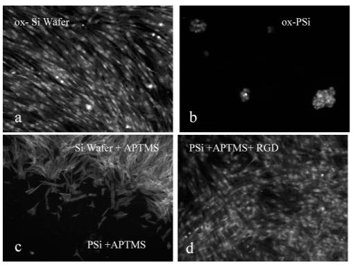
Figure 6.

Fluorescence microscopy images of a dermal fibroblast after 48 h of culturing in 10% FBS—DMEM: (a) smooth silicon wafer (Θc < 10°) with oxide and a PI stain; (b) thermally oxidized PSi (Θc <10°) with a PI stain; (c) oxidized and APTMS-treated PSi—wafer interface with an actin FITC stain; and (d) RGD—peptide-modified PSi with a PI stain. Samples were placed in a 12-well plate, and each well was seeded with ∼1 × 10⁴ cells/cm2 and incubated for 48 h.
