
Figure 5.
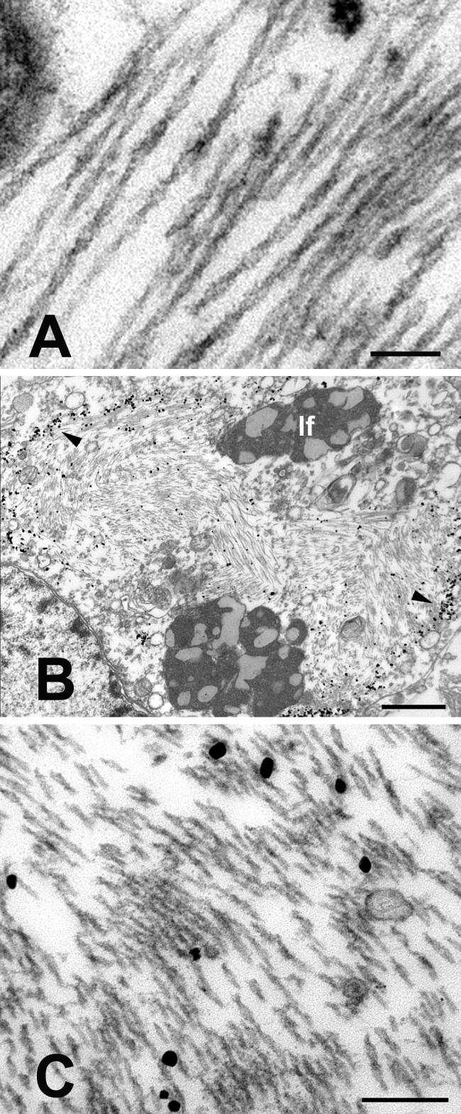
Ultrastructure of paired helical filaments in neurons from the left prefrontal cortex of the aged chimpanzee. A: High magnification view of intraneuronal paired helical filaments. The twisting, filamentous ribbons have a mean half-periodicity of ∼79nm, a maximum width of ∼20nm and a minimum width of ∼10nm. B-C: CP13/immunogold-labeled, intraneuronal paired helical filaments. B is a low magnification view of a CP13-immunoreactive neuron; the nucleus is to the lower left. Because the thick section was immunostained prior to sectioning, the immunogold preferentially decorates the periphery of the mass of PHFs (arrowheads). Note the heavy bundles of paired helical filaments in the cytoplasm. C shows a higher magnification view of CP13/immunogold-labeled PHFs from the cell in B. lf: lipofuscin. Bars = 100nm (A), 1μm (B) and 200nm (C).