
Figure 3. ECDs in G4D-C57 late gestation embryos.
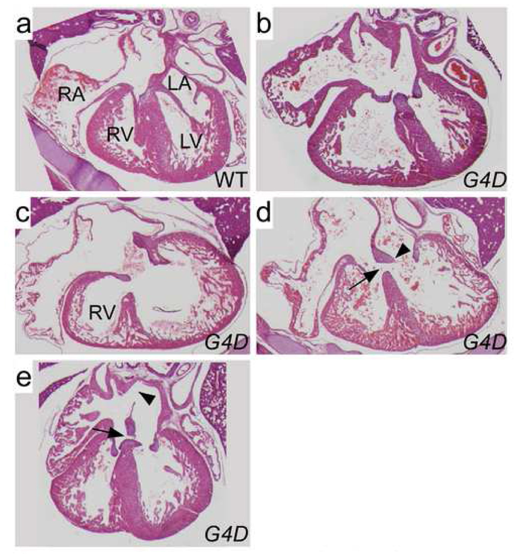
a–e. Hematoxylin and eosin stained sections demonstrating a spectrum of ECDs. a. WT control. b. Well-balanced CAVC canal defect. c. CAVC defect opening mainly into the left ventricle. The RV is moderately hypoplastic. d. Inlet VSD. The atrial septum is intact, and there are two AV valves, albeit with highly primitive leaflets (arrowhead). The ventricular portion of the AV canal is not septated, resulting in an inlet VSD (arrow). e. ASD primum (arrow) and ASD secundum (arrowhead). The ventricular portion of the AV canal is septated.