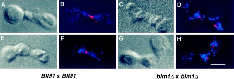
Figure 5.
Microtubule and nuclear morphology in wild-type and mutant zygotes. Wild-type (DBY6654 × DBY7306, A–B and E–F) or bim1Δ strains (DBY7303 × DBY7305, C–D and G–H) were allowed to mate for 3 h and then were fixed for immunofluorescence. Nomarski images (A, C, E, and G) are shown next to combined images of DAPI-stained DNA and antitubulin-stained microtubules (B, D, F, and H). Bar, 4 μm.

