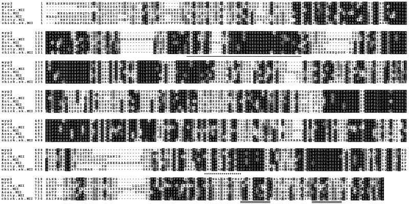
Figure 2.
Sequence alignment of myp2+ with six myosin-IIs from other organisms. The catalytic domain of myp2+ was aligned with myosin-IIs from S. pombe, S. cerevisiae, amoebae, and chicken using Clustal W (Thompson et al., 1994). From top to bottom: S. pombe myp2+, S. pombe myo2+, S. cerevisiae myosin-II (MYO1p), Entamoeba myosin-II, Acanthamoeba myosin-II, Dictyostelium myosin-II, and chicken skeletal muscle myosin-II. The sequence accession numbers are given in MATERIALS AND METHODS. The numbers to the left of the amino acid sequence indicate amino acid position. Amino acid identities are shown in white lettering against black, conserved residues are shown in black lettering against gray. The putative ATP-binding motif is underlined. The putative actin-binding site has asterisks underneath. The putative IQ motifs are underlined twice.

