Figure 4.
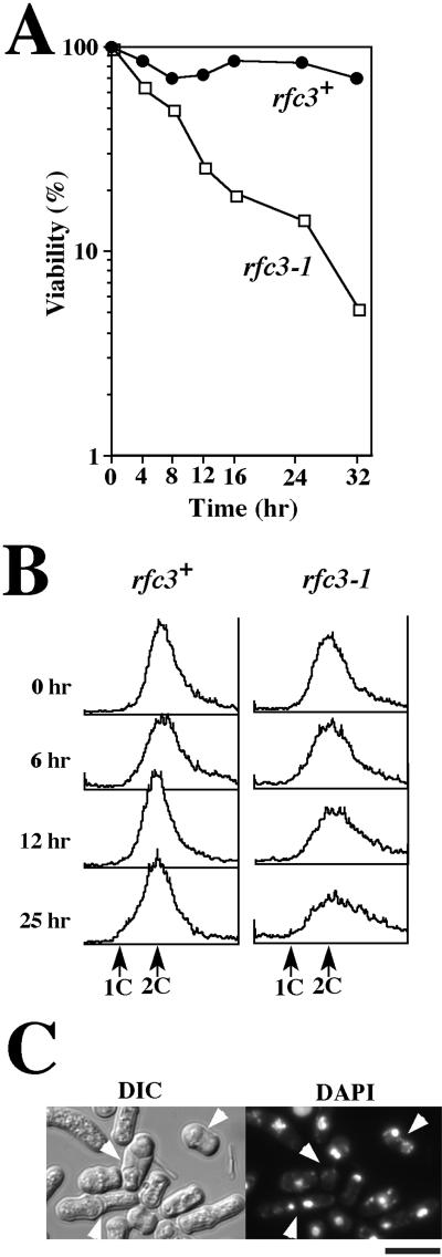
Growth phenotypes of the rfc3-1 mutant. (A) Cell viability of the rfc3+ (MSY10) and the rfc3-1 mutant (MSY11). Exponentially growing cells at 25°C were shifted to incubation at 37°C (time = 0). After 4, 8, 12, 16, 25, and 32 h, a fixed number of cells were removed, diluted, plated onto EMM2 plates supplemented with leucine, and incubated at 28°C for 4 d. Colonies were scored, and viability was expressed as a percentage of the colonies that formed on samples plated immediately before temperature shift. (B) The DNA content of rfc3+ and rfc3-1 cells incubated at 37°C for 0–25 h was estimated by the FACScan method. Positions of the cells with 1C and 2C DNA are shown by arrows. (C) Morphology of cells incubated at 37°C for 32 h. DNA was visualized by staining cells with DAPI. In the rfc3-1 mutant, cells with abnormal nuclear divisions are denoted by arrowheads. Cell morphology was recorded with a charge-coupled device camera. Bar, 10 μm.

