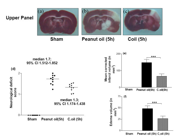
Figure 3.

Protective effect of C.oil on brain infarct and edema volumes and neurological evaluation from rat subjected to middle cerebral artery occlusion at 5 hrs.Upper panel showing the representative photograph of TTC stained coronal brain section after 5 hrs of ischemia from sham-operated (a), peanut oil treated ischemic (b) and ischemia with C.oil post-treatment (c) group at 5 hrs. Unstained areas represent the infarcted brain tissue. Bar graph (d): Neurological evaluation scores of rats from sham-operated, peanut oil treated ischemic and C.oil post-treatment ischemic groups after 5 hrs of ischemia as shown in scattergram. The line demarcates the median. Comparisons were made between C.oil post treated vs. peanut oil treated ischemic group by nonparametric Kruskal-Wallis test followed by Dunn's Multiple Comparison Test. Ten rats/group was used. Bar graph (e): The bar chart showing the infarct volume of brains from sham-operated, peanut oil treated ischemic and C.oil post-treatment ischemic. Data are expressed as mean ± S.E.M. P < 0.001 was considered highly significant when comparisons were made with the peanut oil treated ischemic group by one way ANOVA followed by Newman Keuls post hoc test. Bar graph (f): Effect of post-treatment of C.oil on edema volume after 5 hrs of ischemia. Data are expressed as mean ± S.E.M. of five animals each group. P < 0.001 was considered significant when comparisons were made with the peanut oil treated ischemic group by one way ANOVA followed by Newman Keuls post hoc test.
