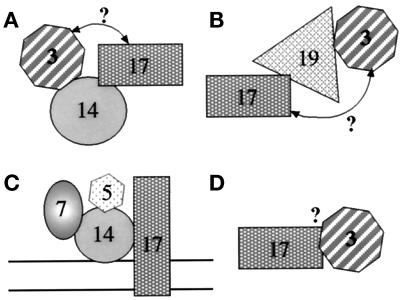
Figure 12.
Schematic representation of various putative Pex protein subcomplexes. The rationale for the definition of these complexes is explained in DISCUSSION. The question mark by the arrows represents a putative bridged interaction, which is not proven or disproven by the data. Because complex formation may occur on the membrane or in the cytosol, no membrane bilayer was included in the diagram for A, B, and D.

