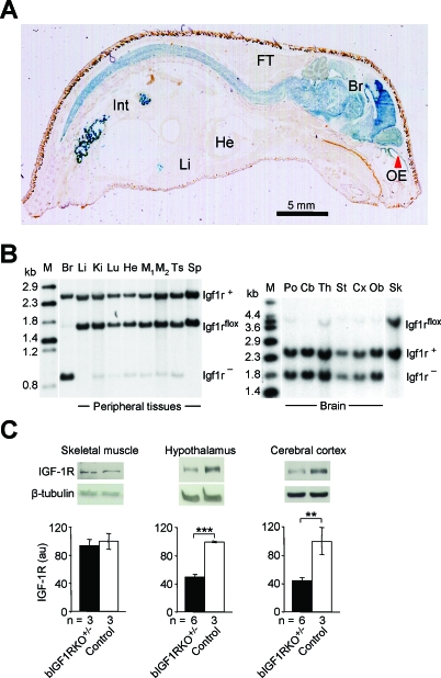
Figure 1. Brain-Targeted Inactivation of the Igf1r Gene Using Cre-lox Mutagenesis.
(A) CNS-specific Cre-lox recombination demonstrated using X-Gal staining (blue) in a sagittal section from a 2-wk-old NesCre +/0 mouse harboring a LacZ reporter (Rosa26R +/0) [33]. NesCre is expressed in neuroepithelium by neuronal and glial precursors. Abbreviations: Br, brain; FT, fat tissue; He, heart; Int, intestine (with bacterial artifacts); Li, liver; OE, olfactory epithelium (red arrow).
(B) Southern blot analysis of adult bIGF1RKO +/− tissues revealed complete recombination in the brain (Br) and the intact Igf1rflox allele in all peripheral tissues (left panel). Recombination in peripheral tissues was minimal. The IGF-1R knockout was effective throughout the brain (right panel) and stable through time (unpublished data). The restriction enzymes used were HincII and I-SceI (left blot) and HincII alone (right). Cb, cerebellum, Cx, cortex, Ki, kidney, Lu, lung, M, DNA size marker, M1/M2, skeletal muscle, Ob, olfactory bulb, Po, pons, Sk, skin, Sp, spleen, St, striatum, Th, thalamus, Ts, testis.
(C) bIGF1RKO +/− mice had normal IGF-1R levels in peripheral tissues (e.g., muscle) and ∼50% of normal levels in the CNS (here: hypothalamus and cortex), as assessed by western blotting.

