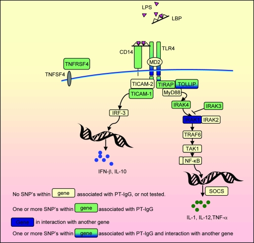
Figure 1. Summary of the TLR pathway in antigen-presenting cells and the main results of this paper.
TLRs recognize molecular patterns associated with a broad range of pathogens including bacteria, fungi, protozoa and viruses. Vaccine components in WCV vaccine that may be recognized by TLR4 include LPS and PT. Following TLR4 activation, both the MyD88 and TICAM1 routes, leading to the expression of proinflammatory cytokines and type I IFNs respectively, may be activated. These promote the development of helper T cell responses providing T-cell help to B cells. TLR signaling in B cells may further promote the generation of antibody responses and the maintenance of serologic memory. TNFRSF4 is expressed on activated T cells. Adapted from BioCarta (http://www.biocarta.com/pathfiles/h_tollPathway.asp), KEGG (Kyoto Encyclopedia of Genes and Genomes) [50], and Metacore™ (http://www.genego.com/metacore.php).

