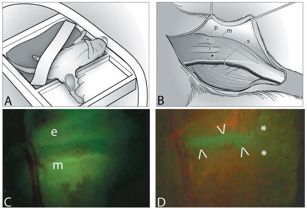
Figure 2.
For surgery and imaging the mouse was positioned in dorsal recumbency, on a heating pad under isoflurane anaesthesia (A). A surgical approach was made to the medial side of the left tibia using the patellar ligament (p), the medial collateral ligament (m) and the saphenous vessels and nerve (s) as landmarks (B). An asterisk (*) indicates the position of the growth plate with the perichondrium intact. The surgical field is illuminated to demonstrate the yellow-green fluorescence of OTC in the epiphyseal (e) and metaphyseal (m) bone (C). The growth plate appears as a non-fluorescent band between the two areas of fluorescing bone. Figure 2D is the identical surgical field, illuminated to show the green fluorescence of the GFP-positive chondrocytes of the growth plate, between the arrowheads. Asterisks mark the medial collateral ligament.

