Figure 3.
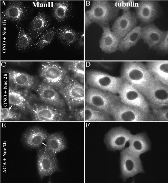
ManII accumulates in an ER intermediate before ministack formation. Clone 9 cells were incubated in 5 μM ONO-RS-082 (A–D) or 25 μM ACA (E and F) at 4°C for 20 min and shifted to 37°C in the continued presence of antagonist plus 6 μg/ml nocodazole for 1 h (A and B) or 2 h (C–F). All conditions included 2 μg/ml cycloheximide. Cells were fixed and processed for double-label immunofluorescence using a polyclonal antibody against ManII (A, C, and E) and a monoclonal antibody against α-tubulin (B, D, and F). The accumulation of ManII in a ring around the nucleus (C and E, arrows) is indicative of its localization in the nuclear envelope and ER.

