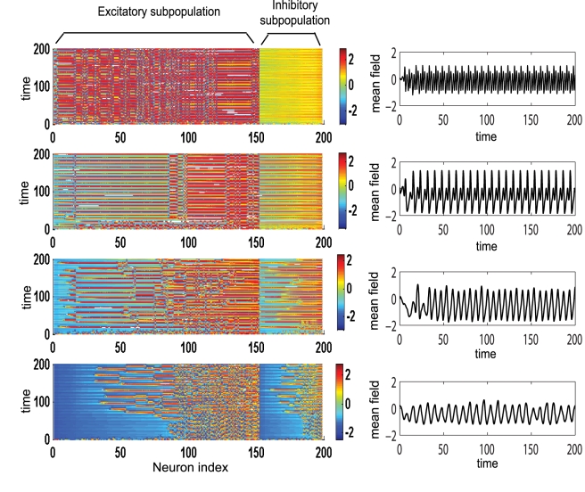
Figure 6. Temporal dynamics of a dominantly inhibitory neural population.
Left: Amplitude color coded time series for all neurons calculated for the following parameter values (starting from bottom to top): K 11 = 0.5 (Region I); K 11 = 0.9 (Region I); K 11 = 2.1 (Region II); K 11 = 3.0 (Region III); for all subfigures n = 1.3, m = 0 and σ = 0.3. Right: the time series of the mean field of the entire population calculated for the same parameters.

