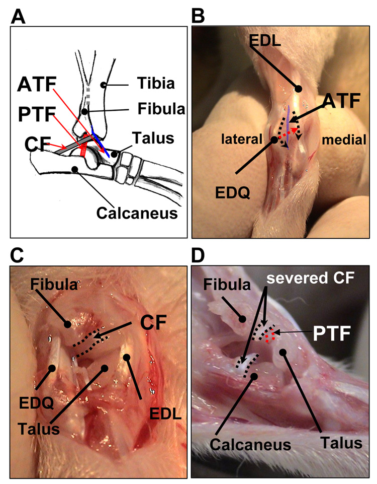
Fig. 1.
(A) A schematic drawing of the lateral ankle ligaments: the anterior talofibular (ATF), the calcaneofibular (CF) and the posterior talofibular ligaments (PTF) (slightly inverted-lateral view). (B, C and D) Surgical procedures for ankle ligament injury (right foot): (B) anterior view, (C) anterolateral view, and (D) lateral view of the ankle.

