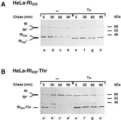
Figure 6.
The truncated ribophorin I variants are stabilized in tunicamycin-treated HeLa cells. HeLa-RI332 (A) and HeLa-RI332-Thr (B) cells were left untreated (lanes a–d and a′–d′) or preincubated with tunicamycin (Tu, 5 μg/ml; lanes e–h and e′–h′). The cells were pulse labeled for 10 min and chased for up to 90 min in the continued absence or presence of the drug. Anti-ribophorin I immunoprecipitations performed on cell lysates were analyzed by SDS-PAGE and fluorography. RI* and RI332* indicate the positions of nonglycosylated endogenous ribophorin I and RI332, respectively, observed in the presence of tunicamycin.

