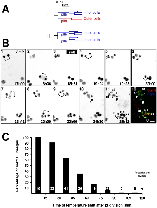
Figure 2. Cells are more receptive to endogenous N-pathway activation during their first hour of life.
The acquisition of a pIIa identity was used as an index of Notch pathway activation in the posterior secondary precursor cell. A Nts/Y; neu>H2B::YFP pupae imaged in vivo was temperature shifted from 30°C to 18°C at a given moment after pI division enabling the N-receptor to be endogenously activated. A) Lineages expected if the posterior secondary precursor cell responded or not to Notch activation (i and ii respectively). (B) Representative frames from an in vivo recording followed by immunostaining of two microchaete lineages. Anterior is on the left. The temperature shift was applied at 18h54 after pupal formation (APF). Brackets indicate cell divisions. Time APF is shown at the bottom right of each frame. At 18°C, development proceeds half as quickly as at 25°C. The arrowhead (frame 10) indicates apoptosis of the glial cell. In frame 12, bristle lineage cells are revealed by anti-GFP antibody (green). Su(H) and Repo immunoreactivities were used to identify socket (so) and glial (g) cells respectively (yellow and blue). The other cell identities have been ascribed based on the characteristics of their divisions recorded in vivo, n: neuron, st: sheath, sh: shaft cell. Each image results from the merge of 5 horizontal optical sections. Note that when the temperature shift was applied 1h54 after the pI division (upper cluster), the resulting organ was formed exclusively by inner cells. In contrast, when applied 0h18 after the pI division (lower cluster), a normal organ resulted. Scale bar: 5 µm. (C) Percentage of normal sensory organs observed when the temperature shift was applied at different moments after pI division (abscissa). Note that normal sensory organs were observed when the temperature shift was applied during the first 45 min of the secondary precursor cells life. In this and other histograms, the numbers at the base of the bars indicate the number of sensory clusters analyzed.

