Figure 5.
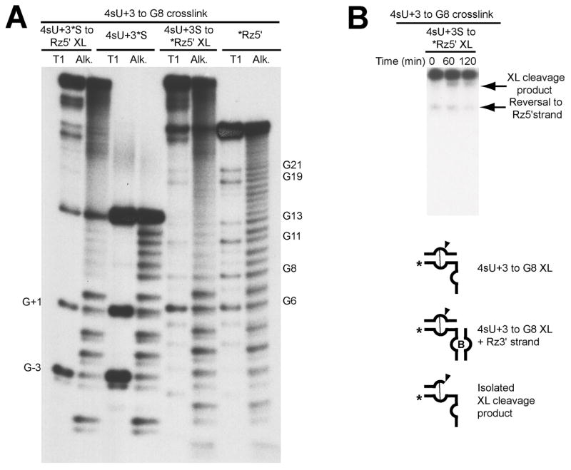
4sU+3 crosslink. A) Mapping of 4sU+3 crosslinked sites. The crosslinked sites were mapped by limited akaline hydrolysis (Alk) and partial digestion with RNAse T1 (T1). The species consisting of 5′-32P end-labeled 4sU+3 substrate crosslinked to ribozyme 5′ strand (4sU+3*S to Rz5′ XL), and the end-labeled 4sU+3 substrate control (4sU+3*S) are paired on the left. The species consisting of 5′-32P end-labeled ribozyme 5′ strand crosslinked to 4sU+3 substrate (4sU+3S to *Rz5′ XL), and end-labeled ribozyme 5′ strand control (*Rz5′) are paired on the right. Sites of T1-induced cleavages are indicated by the numbered guanosine. B) Activity assay for isolated crosslinked species. Ribozyme 3′ strand (Rz3′) was added to crosslinked products,labeled on ribozyme 5′ strand as indicated, in the presence of 1 mM cobalt hexaammine for up to 2 hours. The crosslink cleavage product and the reversal to ribozyme 5′ strand are shown by arrows.

