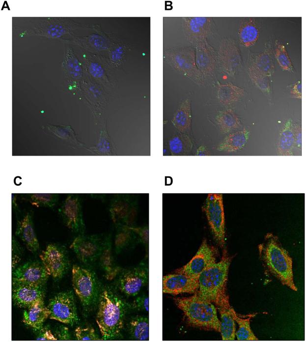
Fig. 4.

Cellular localization of fluorescently labeled DNA and/or Pluronic 123 examined by confocal microscopy on fixed NIH 3T3 cells. (A, B) Cells were transfected for 30 min with Exgen 500-based polyplex without (A) or with (B) 0.03 % Pluronic P123. Signals correspond to YOYO-1 labeled gWIZLuc pDNA (green), RITC-labeled Pluronic P123 (red) and ToPro-3 stained nuclei (blue), respectively. (C, D) Cells were exposed to 0.03 % Pluronic P123 alone for 60 min and then examined for co-localization of Pluronic P123 with caveolin-1 (C) and clathrin (D). Signals correspond to FITC-labeled Pluronic P123 (green), caveolin-1 (red), clathrin (red) and ToPro-3 stained nuclei (blue).
