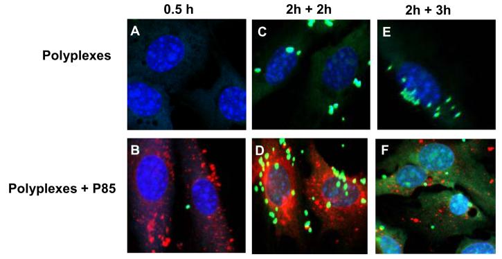
Fig. 5.
Effect of Pluronic on intracellular localization of DNA in NIH 3T3 cells. Confocal microscopy images of cells transfected with Exgen 500-based polyplexes with (B, D, F) or without (A, C, E) 1% Pluronic P85. Cells were exposed to polyplex for (A, B) 30 min or (C-F) 2 h and then incubated for additional 2 (C, D) or 3 h (E, F) in a fresh medium. Signals correspond to YOYO-labeled pDNA (green), RITC-labeled Pluronic P85 (red) and ToPro-3 stained nuclei (blue), respectively. This figure is representative of multiple areas in confocal images.

