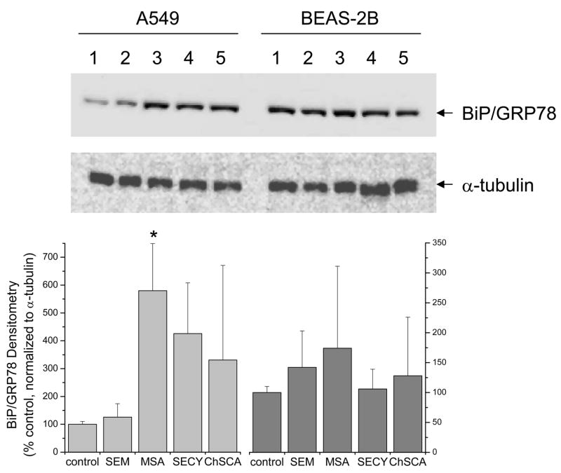
Figure 5. Induction of BiP/GRP78 expression by selenocompounds in human lung cells.
Cell lysates were homogenized and 10 μg of protein was loaded onto a NuPAGE 10% Bis-Tris gel and transferred to a PVDF membrane for immunochemical analysis as described in the Methods. Cells were incubated with the selenocompounds for 24 hrs at the same concentrations indicated in figure 4. Immunochemical analysis of α-tubulin was utilized as a loading control. Immunochemical analysis of BiP/GRP78 was evaluated as an indicator of the unfolded protein response. A representative blot of triplicate experiments is shown in the upper panel and densitometry for all experiments is shown in the lower panel with MSA demonstrating a significant difference compared to the control (*, P<0.05).

