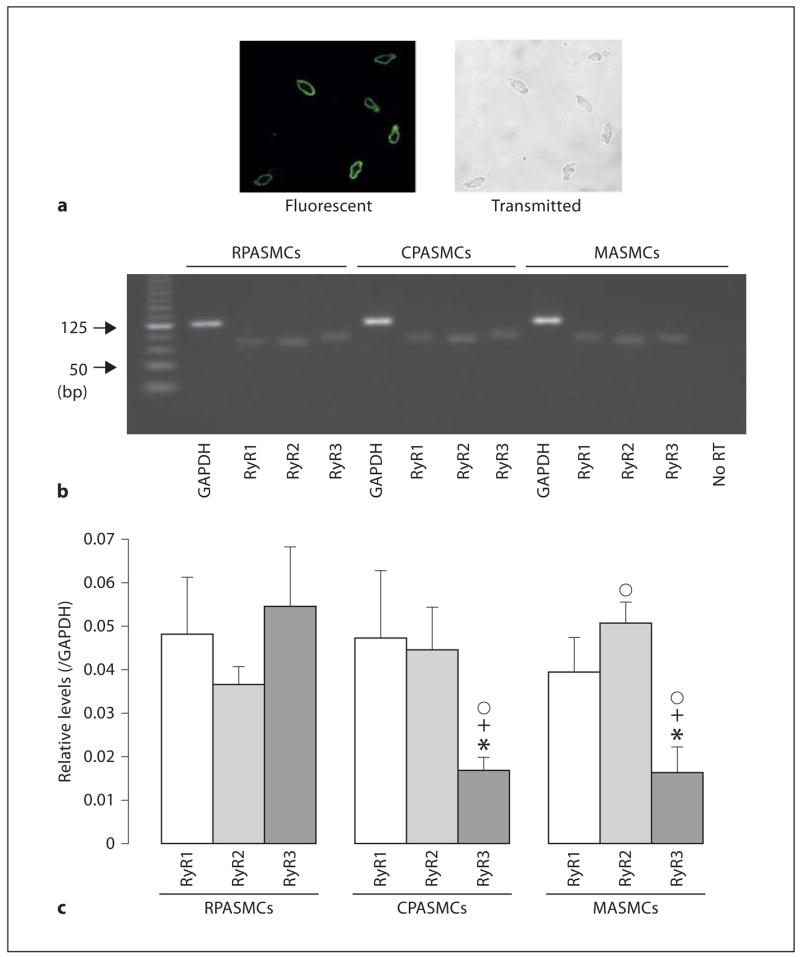
Fig. 1.

RyR subtypes are heterogeneously expressed in freshly isolated mouse RPA-SMCs, CPASMCs and MASMCs. a Expression of the smooth muscle-specific actin was found in nearly all isolated cells from resistance pulmonary arteries. Fluorescence image for actin staining (left) and transmitted light image (right) were simultaneously taken using a Zeiss LSM510 laser scanning confocal microscope. Cells were incubated with a primary antibody for smooth muscle-specific actin and then stained with Alexa Flour 488-conjugated anti-mouse antibody. b Gel electrophoresis reveals expression of RyR1, RyR2 and RyR3 mRNAs in RPASMCs, CPASMCs and MASMCs. The predicted PCR product sizes for RyR1, RyR2, RyR3 and GAP-DH were 81, 77, 83 and 122 bp, respectively. c Graphs show the relative mRNA expression levels of RyR1, RyR2 and RyR3 in RPASMCs, CPASMCs and MASMCs. The relative mRNA expression levels were obtained by normalizing the absolute expression levels of RyR1, RyR2 and RyR3 to that of GAPDH. Data were obtained from 6 separate experiments. * p < 0.05 compared with RyR1 in the same type of vascular SMCs; + p < 0.05 compared with RyR2 in the same type of cells; °p < 0.05 compared with RyR2 or RyR3 in RPASMCs.
