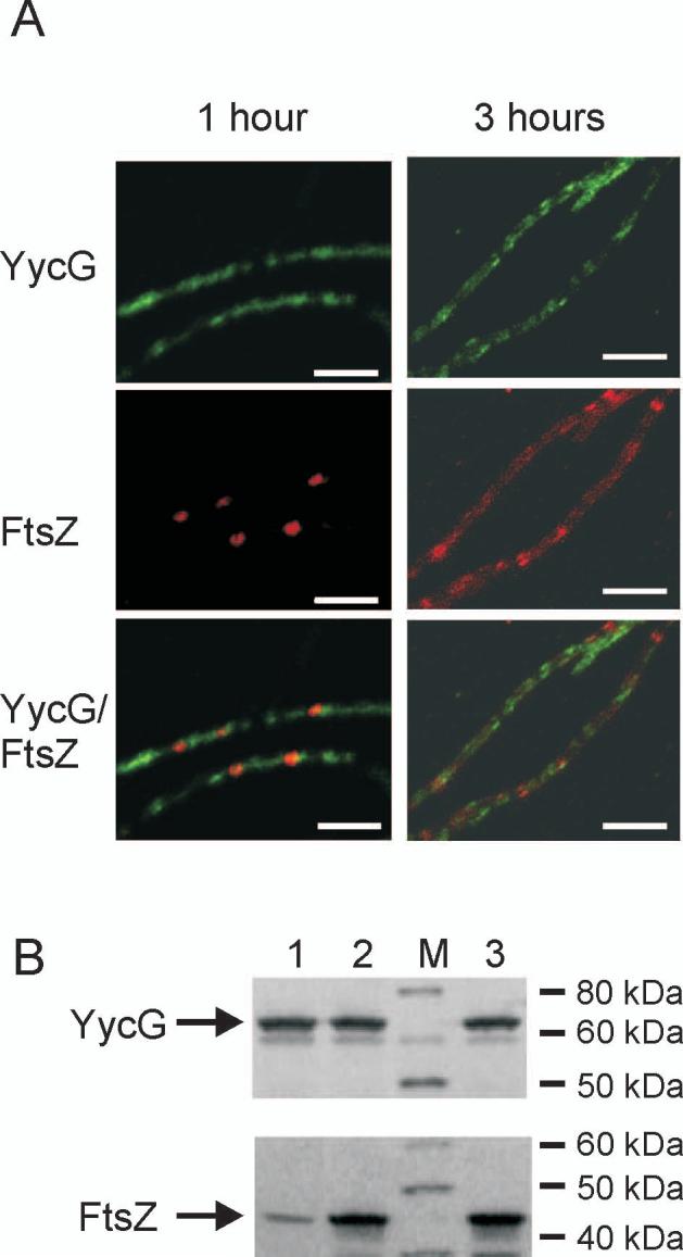
Figure 2.

YycG does not localize to the septum in the FtsZ-depleted strain. (A) YycG (green) and FtsZ (red) were visualized immunologically in KP444 cells following 1 and 3 hours growth at 37°C in the absence of IPTG to repress FtsZ expression. Bars indicate 5 μm. (B) Cellular protein levels of YycG and FtsZ were visualized by western blotting using whole cell protein extract derived from strain KP444 grown for one hour either in the absence (lane 1) or presence of 1 mM IPTG (lane 2) or from wild type strain JH642 (lane 3) as reference; Lane M, molecular weight standard.
