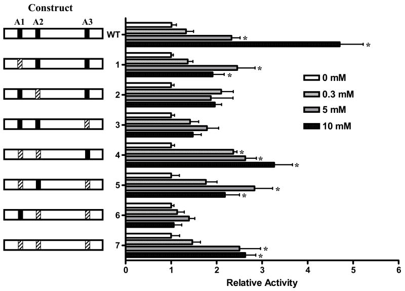
Figure 5. Mutagenesis of A boxes impairs glucose-responsiveness of the PANDER promoter.
(A) Mutagenized PANDER/luciferase plasmids are shown on the left side of the above figure and denoted by construct number as described previously (Figure 2A). A boxes are designated as A1, A2, and A3. Intact A boxes (5′-TAAT-3′) are represented by a black bar whereas mutagenized sites (5′-TAGT-3′) are shown by hatched bars. PANDER/luciferase constructs were transiently co-transfected with Renilla luciferase reporter plasmid phRL-TK into βTC3 cells in glucose-free DMEM. Four hours following transfection, the medium was replaced with DMEM containing incremental concentrations of D-glucose (i.e., 0 mM (white bar), 0.3 mM (vertical hatched bar), 5 mM (gray bar), 10 mM (black bar)). Twenty-four hours post-transfection, luciferase activity was detected by luminometry. Luciferase activity is shown as fold above respective construct at 0 mM glucose and normalized to renilla expression of the phRL-TK plasmid. All data are shown as mean ± S.E. from three independent experiments with each construct evaluated in triplicate. Asterik indicates a P value < 0.05 for the comparison of the PANDER/luciferase construct with that of the activity at 0 mM glucose according to ANOVA.

