
Fig. 8. Lopinavir (LPV) stimulates activity of AMPK.
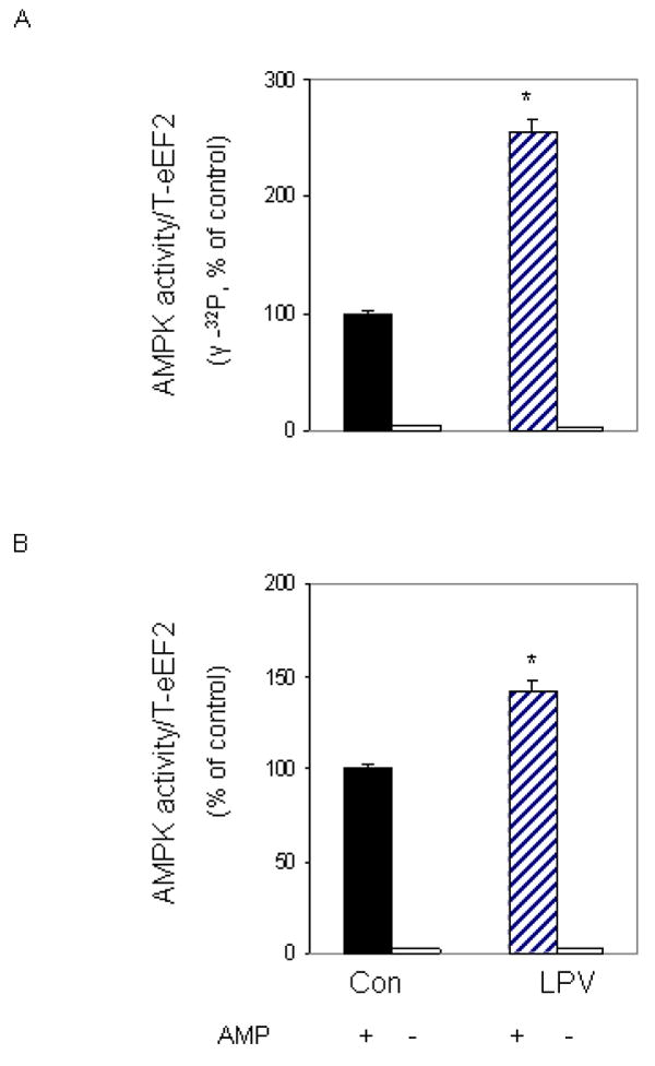
C2C12 myocytes were incubated in the presence or absence of LPV (10 μM) for 15 min. For in vitro kinase activity, AMPK was immunoprecipitated from 100 μg of cell lysates and the activity was assayed using eEF2 as the substrate, while in the presence of MgCl2 and AMP. Reaction mixtures were incubated in the presence (panel A) or absence (panel B) of [γ-32P] ATP as described under “Materials and Methods”. In panel B, reaction mixtures were examined by Western blot, using the anti-phospho eEF2 (T56) antibody. Results are mean ± SE of 3 independent experiments consisting of 4 replicate samples per experiment. * P<0.05 versus control values.