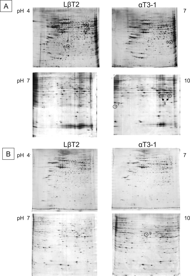
FIG. 1.
Two-dimensional gel electrophoresis of proteins from isolated nuclei (A) or solubilized nuclear proteins (B) of untreated mature LβT2 and immature αT3–1 gonadotrophs over the pI ranges 4–7 or 7–10. Nuclei were prepared as described in the Materials and Methods, before resolving over first and second dimensions. Numerous pairs of samples were run and compared, and representative pairs are shown. Protein spots showing differential expression patterns that were isolated for identification are numbered; protein 8 appears in two distinct spots on the gel (as revealed by its subsequent identification).

