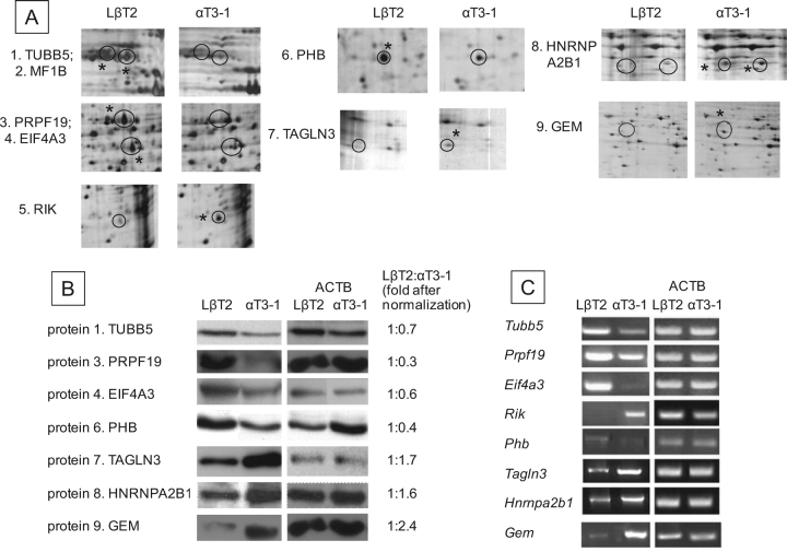
FIG. 2.
Identification and verification of differentially expressed proteins. A) The differentially expressed proteins marked in Figure 1 are shown magnified; an asterisk marks the spot of higher intensity for each pair. B) After the proteins were excised, in-gel digested, and subjected to mass spectrometry, a putative identity was obtained and, where antisera were available, Western analysis was carried out to verify the identification and examine differential expression in nuclear extracts of the two cell lines, with ACTB as internal control. The expression levels of these proteins were assessed, and are shown, after normalization, relative to levels in LβT2 cells. C) RT-PCR was also carried out to determine whether the differential protein levels relate to differential gene expression, with Actb as internal control. The 1110005A23RIK is abbreviated to RIK in all figures.

