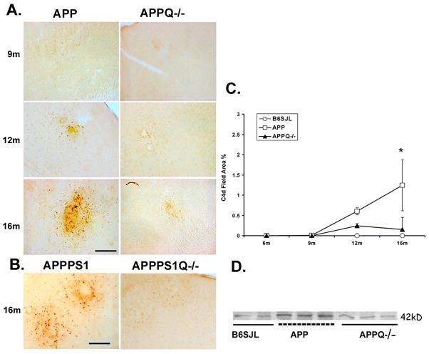
Figure 2. APPQ−/− have less C4 reactivity than APPQ+/+ mice.
A. Representative pictures of cortex of APPQ+/+ (left) and APPQ−/− (right) mice at different ages showing immunostaining with anti-mouse C4 (C4d) (HBT). Scale bar: 50μm. B. Sections from cortex of 16 mo old double transgenic APPPS1 Q+/+ and APPPS1Q−/− were stained with anti-mouse C4 as in A. C. Quantification of C4 reactivity by image analysis shows that C4d staining in APPQ+/+ (-□-) mice is significantly higher (* p<0.02) than in APPQ−/− (-▲-) at 16 mo. (n=3 or 4 mice for each genotype at each age). There was no reactivity in the wild type B6/SJL (-○-). Data points represent group means ± SD. D. Western blot analysis of brain extracts from 16m B6SJL, APPQ+/+ and APPQ−/− mice probed with polyclonal anti-mouse C4 (Ogata, Table 1) detect C4 activation fragment, C4d at 42,000 Mr.

