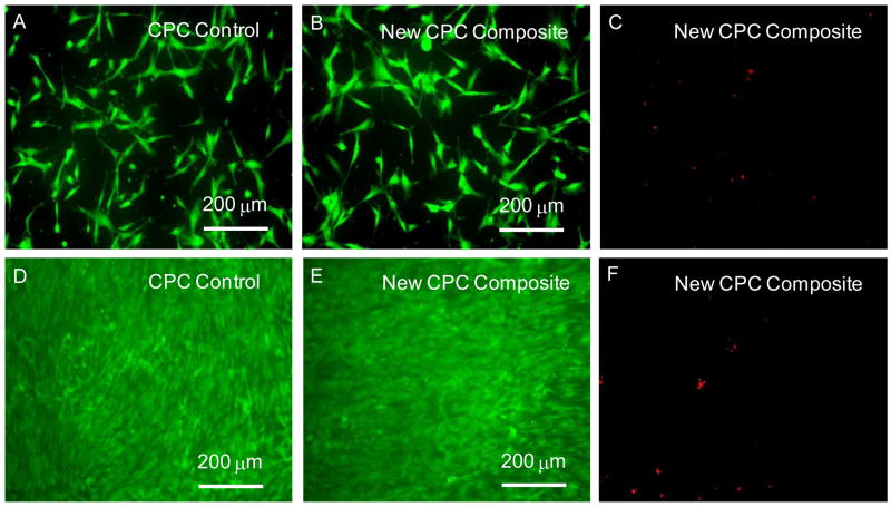
Figure 4.
(A–C) After 1-day culture, live cells (stained green) appeared to have adhered and attained a normal polygonal morphology on the specimen. Dead cells (stained red) in (C) were very few on both materials. (D–F) After 14 days of culture, live cells had formed a confluent monolayer on all specimens. The cell density was much greater than the 1-d density, indicating that the cells had greatly proliferated. These results suggest that cell proliferation was similar, demonstrating that the new CPC composite was as non-cytotoxic as the FDA-approved CPC control and the TCPS control.

