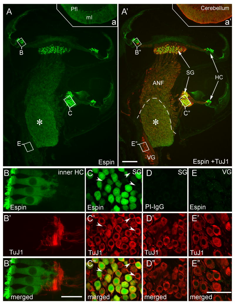
Fig 1.
Immunofluorescence localization of espins in cryosections of adult rat cochlea. Sections of decalcified inner ear labeled with affinity purified rabbit polyclonal espin antibody or preimmune IgG (PI-IgG) (green) and TuJ1 class III β-tubulin antibody (red). A,A′: Intense espin antibody labeling is present in hair cells (HC), in neurons of the spiral ganglion (SG) and in their auditory nerve fibers (ANF). Neurons of the vestibular ganglion (VG) are not labeled with espin antibody. The Schwann-glia border is shown with a dashed line in A′, and the asterisk in A,A′ marks the central nervous system portion of the ANFs, which shows elevated espin antibody labeling. A portion of the cerebellar parafloccular lobule (Pfl) that was processed together with the cochlear tissue in the temporal bone is shown in insets a,a′. Espin antibody labels the cerebellar Purkinje cells and their dendritic arbors in the molecular layer (ml) (Sekerková et al., 2003). B-B″: Espin antibody does not label the peripheral processes of the SGNs, which connect to the intensely espin-positive inner hair cells (inner HC). C-C″: Espin antibody labels a large proportion of SGNs. Arrowheads point to examples of espin-negative, but TuJ1-positive type-I SGNs. D-D″: Spiral ganglion from a neighboring section showing no labeling when preimmune IgG (PI-IgG) is substituted for espin antibody. E-E″: Espin antibody does not label the vestibular ganglion (VG). Scale bars, A-A′, 200 μm; B-B′, 20 μm: C-E″, 50 μm. A magenta-green version of this figure can be viewed online as Supplementary Figure 1.

