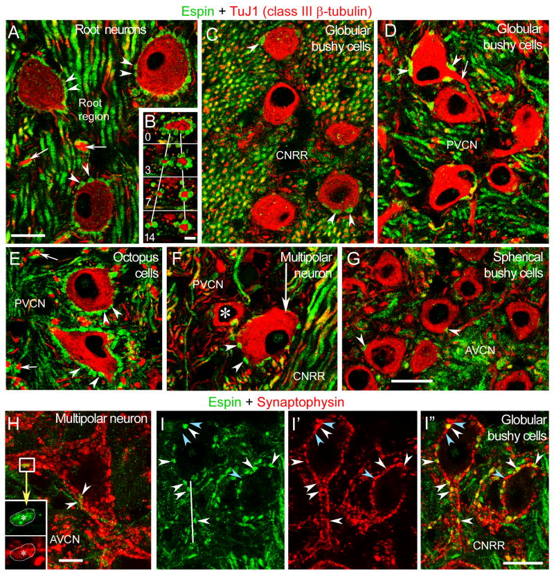
Fig. 5.
Confocal immunofluorescence images from various CN regions labeled with espin antibody (green) and either TuJ1 class III β-tubulin antibody (red) (A-G) or synaptophysin antibody (red) (H-I″). A-G: These panels illustrate the presence of espin in synaptic boutons (arrowheads in A-G) that contact a variety of cells and dendrites (B and arrows in A,D,E). B, images of an emerging dendrite surrounded by espin-positive boutons taken at 0, 3, 7, and 14 μm focusing intervals. Asterisk in F, globular bushy cell. CNRR, central nerve root region. H: A large multipolar neuron receives only a few espin-positive boutons (arrowheads and inset). This is a stack of 6 consecutive 1 μm thick confocal images. I-I″: Espins colocalize with synaptophysin in boutons contacting globular bushy cells (white arrowheads). This is a stack of 2 consecutive 1 μm thick confocal images. Blue arrowheads point to examples of espin-negative, synaptophysin-positive boutons. CNRR, central nerve root region. Scale bars: A, C-I″, 20 μm; and B, 5 μm. A magenta-green version ofthis figure can be viewed online as Supplementary Figure 4.

