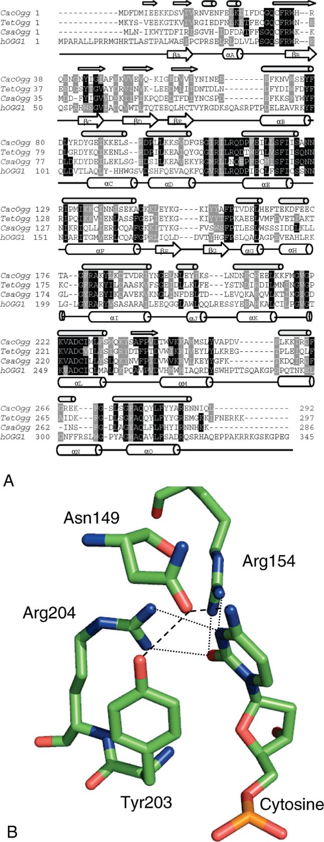
Figure 2.

(A) Sequence alignment of CacOgg, hOGG1, and putative Oggs from Thermoanaerobacter ethanolicus (Tet) and Caldicellulosiruptor saccharolyticus (Csa). The PSIPRED prediction (36) of CacOgg secondary structure is indicated by small arrows ( β-sheets) and small cylinders (α-helices) directly above the CacOgg sequence. The hOGG1 secondary structure (18) is indicated by large arrows and cylinders drawn under the hOGG1 sequence. (B) Model of hOGG1 interactions with the base opposite the lesion as redrawn from the crystal structure (1EBM). Both dashed and dotted lines indicate H-bonding.
