
Fig. 3.
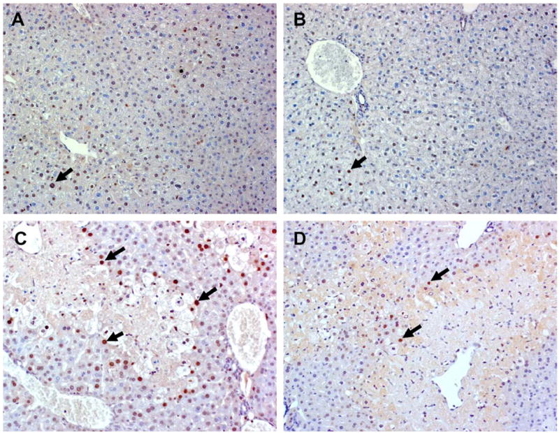
Representative photomicrographs of PCNA immunostaining in hepatocyte nuclei of WT and JNK2−/− mice 24 h after treatment with saline vehicle or APAP. (A) Liver from a saline-treated WT mouse exhibited minimal staining and as did the liver from a saline-treated JNK2−/− mouse (B). (C) Liver from an APAP-treated WT mouse showed a increased number of PCNA positive stained S-phase cells at the periphery of a necrotic lesion, while much less staining was seen in cells surrounding the necrotic lesion in the liver of a JNK2−/− mouse (D). Solid arrows point to PCNA positive S-phase cells, which are characterized by intense brown nuclear staining. Original magnification: 20×.