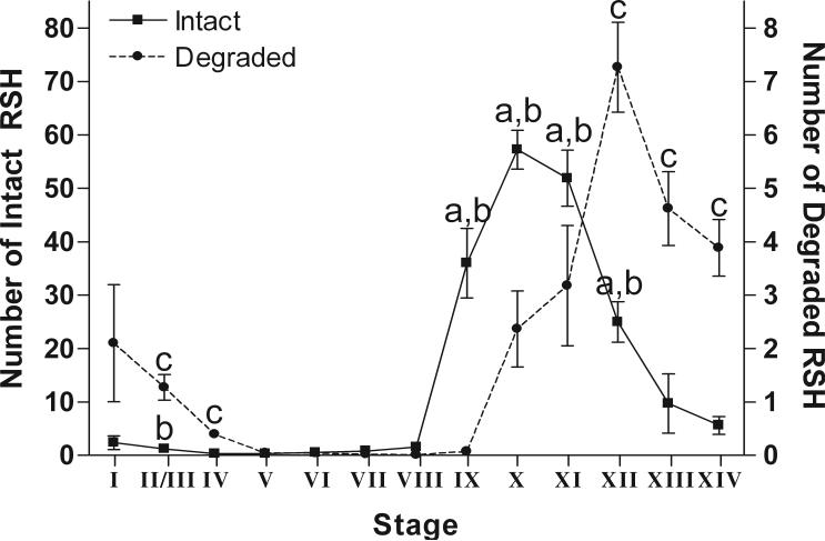
Figure 5.
The average number of intact RSH (left y-axis) or degraded RSH (right y-axis) per seminiferous tubule at each stage from rats treated with 1% HD for 18d. The average number of intact RSH increases soon after spermiation and the average number of degraded RSH increases one stage later. Intact RSH are significantly different from degraded RSH in stages IX-XII (indicated by “a”). There are significantly more intact RSH at stages IX-XII and stage II/III in treated testes compared to control, (indicated by “b”). There is a significantly higher average of degraded RSH in treated versus control testes at stages XII to IV, excluding stage I (indicated by “c”). There is a 10-fold difference between the scale of intact and degraded RSH. Error bars indicate SEM.

