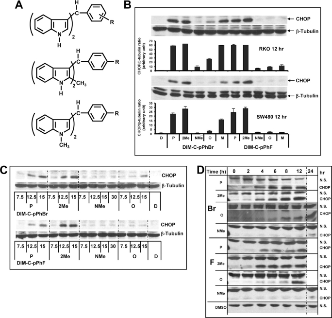
Fig. 2.
Structure-dependent activation of CHOP by DIM-C-pPhBr and DIM-C-pPhF analogs. (A) Structure of DIM-C-PhBr and DIM-C-PhF analogs. Structure-dependent (B), dose-dependent (C) and time-dependent (D) activation of CHOP in colon cancer cells. Cells were treated with either DMSO (D) or various C-DIM compounds at 15 μM for 12 h and changes in protein expression were determined by western blot analysis as described in Materials and Methods. Protein levels were measured with Image J and normalized to β-tubulin or compared with a non-specific (NS) band (Figure 2D). CHOP was non-detectable in cells treated with DMSO (Figure 2D).

