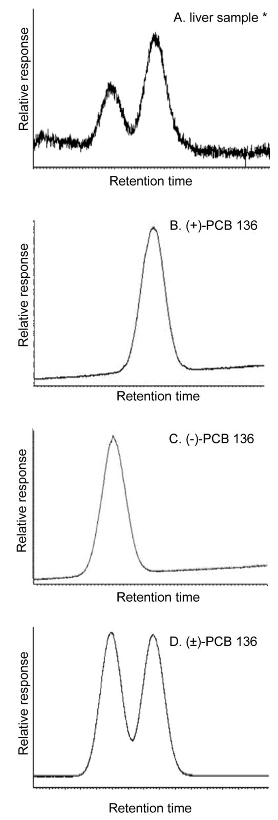
Figure 1.

Separation of PCB 136 atropisomers using chiral gas chromatography. Representative chromatogram illustrating the enrichment of (+)-PCB 136 in the liver of a PB-pretreated mouse (22) (A) as well as chromatograms of pure (+)-PCB 136 atropisomer (B), pure (-)-PCB 136 atropisomer (C) and racemic PCB 136 standard (D) used in the spectral binding studies. The PCB atropisomers were separated using two serially connected Nucleodex P-PM columns as described in Experimental Procedures.
