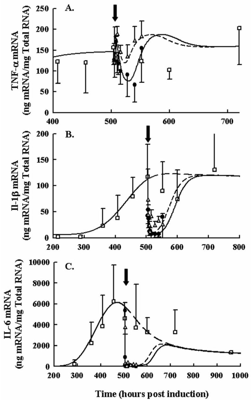
Figure 3.

Time course of TNF-α (Panel A), IL-1β (Panel B), and IL-6 (Panel C) paw mRNA expression in arthritic rats following 0.225 and 2.25 mg/kg SC DEX. Closed circles depict mRNA after single high dose administration. Open triangles depict mRNA after single low dose administration. Black arrows indicate time of dosing, 504 hours post CIA induction. Open squares show natural disease progression of cytokine mRNA. Observations in all figures are reported as mean ± one standard deviation. Model fittings are presented as solid lines for the 2.25 mg/kg dose, short-dashed lines for the single 0.225 mg/kg dose, and long dashed lines for no DEX treatment.
