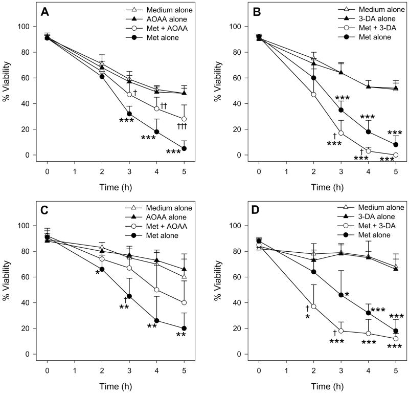
Figure 6.
Time course of the effect of 0.2 mM AOAA (n=4) and 0.1 mM 3-DA (n=3) on the cell viability of male hepatocytes exposed to medium alone or medium spiked with 30 mM Met as determined by TB exclusion (A, B) and LDH leakage (C, D). The symbol * indicates values that were significantly lower than cells incubated with medium alone (*p<0.05, **p<0.01, ***p<0.001). The symbol † indicates values that were significantly different than cells incubated with Met alone († p<0.05, † † p<0.01, † † † p<0.001).

